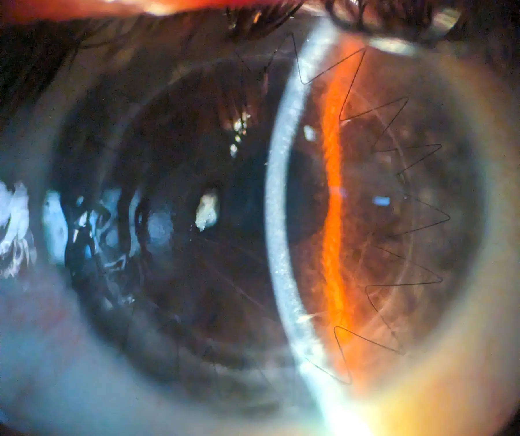
DMEK
DMEK
Descemet membrane endothelial keratoplasty
The cornea, the clear front window of the eye, is essential for sharp vision. Its innermost layer, the endothelium, maintains corneal clarity and proper hydration. When these endothelial cells become damaged or dysfunctional — often through Fuchs’ dystrophy or complications of previous eye surgery — the cornea swells and clouds, significantly impairing vision.
For many years, treating such conditions involved replacing the entire thickness of the cornea (penetrating keratoplasty) or, more recently, a partial-thickness posterior transplant such as DSAEK. While effective, these procedures carry inherent complexities.
DMEK represents a further evolution in corneal transplantation, offering a highly selective approach. Instead of replacing a substantial portion of corneal tissue, DMEK transplants only the diseased Descemet’s membrane and its attached endothelial cell layer. This donor tissue is exceptionally thin — typically around 15–20 microns.
Key aspects and advantages
- Targeted replacement. The core principle is to replace only the compromised cellular layer. This preserves the patient’s own healthy corneal stroma and anterior layers, leading to a more anatomically natural result.
- Faster visual recovery. By minimising alterations to the cornea’s overall architecture, DMEK often allows quicker visual rehabilitation than older techniques. Patients may notice improvement relatively quickly, with the potential for excellent visual acuity.
- Reduced rejection rates. Because a smaller amount of donor tissue is transplanted, and it sits in a more immunologically favourable environment, the risk of graft rejection is generally lower than with full-thickness or thicker lamellar grafts.
- Improved refractive stability. DMEK typically induces less change in corneal curvature, because the incisions are small and the bulk of the patient’s cornea remains untouched. This leads to more predictable refractive outcomes.
- Preserved structural integrity. Leaving the majority of the native cornea intact helps maintain the eye’s structural strength.
Who is a candidate
DMEK is primarily indicated for disease affecting the corneal endothelium where the rest of the cornea is relatively healthy. The most common indications are:
- Fuchs’ endothelial corneal dystrophy
- Pseudophakic bullous keratopathy — corneal oedema after cataract surgery
- Other causes of endothelial failure
A comprehensive ophthalmic evaluation is necessary to determine whether DMEK is the most appropriate surgical option for an individual patient.
The procedure
DMEK is a sophisticated microsurgical procedure. The surgeon carefully removes the patient’s diseased Descemet’s membrane and endothelium. The thin donor graft is then inserted into the eye, unfurled, and positioned against the back of the patient’s cornea. An air or gas bubble is used to hold the graft in place while it adheres.
After surgery
Patients are usually asked to maintain a supine, face-up position for a period after surgery, so the bubble effectively supports the graft. Adherence to post-operative instructions is crucial for a successful outcome.
DMEK has significantly advanced the treatment of corneal endothelial disorders. It offers a less invasive method with the potential for excellent visual outcomes and a quicker recovery for appropriately selected patients — part of the ongoing move in ophthalmic surgery towards more precise and selective solutions.
Sources
Updated: